58 Top Pictures Cat Bone Structure Diagram - Femur bone structure Royalty Free Vector Image. The diaphysis and the epiphysis. Under the microscope, bone can be divided into two types: The brachial artery and median nerve of the cat pass through this structure and are, therefore, somewhat protected by this arch of bone. Vestibulocochlear nerve and sensory structures. The basic microscopic unit of bone is an osteon (or haversian system).
ads/bitcoin1.txt
(b) in this micrograph of the. Odkryj human bone anatomy long bone structure stockowych obrazów w hd i miliony innych beztantiemowych zdjęć stockowych, ilustracji i wektorów w kolekcji shutterstock. You can see these tissues in the diagram above. Osteons are roughly cylindrical structures that can measure several millimeters long and around 0.2 mm in diameter. Structure and physical properties.—bone is one of the hardest structures of the animal body;
Long Bone Anatomy | Human Anatomy Quiz - Quizizz from media.quizizz.com This is the upper long bone of the leg. Cat skeletal anatomy poster created using vintage images. Structure and physical properties.—bone is one of the hardest structures of the animal body; Cheek bone (zygoma) upper jaw (maxilla). Lower jaw (mandible) collar bone. Their unique cat bone structure allows them to twist, turn, and leap, making them graceful and able to perform remarkable feats. An equilibrium between osteoblasts and osteoclasts maintains. Odkryj human bone anatomy long bone structure stockowych obrazów w hd i miliony innych beztantiemowych zdjęć stockowych, ilustracji i wektorów w kolekcji shutterstock.
It is lighter, less dense, and more flexible than compact bone.
ads/bitcoin2.txt
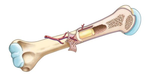
It's time to look inside our feline friends and of course, it might be a bit easier if we know what bone goes where, so let's take a look at the skeleton structure of cats with a useful diagram. The structure of bone with diagram and definitions. The structure of a long bone allows for the best visualization of all of the parts of a bone (figure 1). Bones are made of active, living cells that are busy growing, repairing themselves, and communicating. Irruption of the subperiosteal tissue. The structure of bone marrow constitutes of hematopoietic tissue islands and adipose cells surrounded by vascular sinuses interspersed within a meshwork of trabecular bone. Structure and physical properties.—bone is one of the hardest structures of the animal body; Excessive draft work imposes strain on the hind legs that provide forward locomotion, also requiring. Protection of the heart , lungs , and other organs and structures in the chest creates a problem somewhat. It is also called osseous tissue or cortical bone and it provides structure and support for an organism as part of its skeleton, in addition to being a location for the storage of minerals like. An equilibrium between osteoblasts and osteoclasts maintains. Diagram of the skeleton of a cat. Compact bone tissue forms the outer shell of bones.
It is also called osseous tissue or cortical bone and it provides structure and support for an organism as part of its skeleton, in addition to being a location for the storage of minerals like. Bone basics and bone anatomyhave you ever seen fossil remains of dinosaur and ancient human bones in textbooks, television, or in person at a museum? The basic microscopic unit of bone is an osteon (or haversian system). Under the microscope, bone can be divided into two types: Watch as aaron creates a tiger in photoshop then creates the underlying bone structure.
femur bone diagram - Google Search | Femur bone, Skull and ... from i.pinimg.com Cumulative diagram showing the percentual contribution of skeletal elements to ali pathologicai bones. Cheek bone (zygoma) upper jaw (maxilla). It is also called osseous tissue or cortical bone and it provides structure and support for an organism as part of its skeleton, in addition to being a location for the storage of minerals like. With 30 exercises covering all body systems, a clear, engaging writing style, and full color illustrations, this updated edition offers students everything needed for a successful lab experience. Cat skeletal anatomy poster created using vintage images. The axial skeleton and the. Osteone life spans and the time it takes to produce an osteone vary from species to species and clade to clade. Osteons are roughly cylindrical structures that can measure several millimeters long and around 0.2 mm in diameter.
You can see these tissues in the diagram above.
ads/bitcoin2.txt
The functions of the skeleton are to provide support, give our bodies shape, provide protection to other systems and organs of the body, to provide attachments for muscles, to produce movement and to. Excessive draft work imposes strain on the hind legs that provide forward locomotion, also requiring. What causes the bone disease rickets? Under the microscope, bone can be divided into two types: The diaphysis and the epiphysis. An equilibrium between osteoblasts and osteoclasts maintains. Cancellous (trabecular or spongy) bone: Bones are made of active, living cells that are busy growing, repairing themselves, and communicating. It's time to look inside our feline friends and of course, it might be a bit easier if we know what bone goes where, so let's take a look at the skeleton structure of cats with a useful diagram. The bones of the chest — namely the rib cage and spine — protect vital organs from injury, and also provide structural support for the body. The structure of bone marrow constitutes of hematopoietic tissue islands and adipose cells surrounded by vascular sinuses interspersed within a meshwork of trabecular bone. It consists of a very hard (virtually solid) mass of bony tissue arranged in concentric layers (haversian systems). Diagram of the skeleton of a cat.
Bone basics and bone anatomyhave you ever seen fossil remains of dinosaur and ancient human bones in textbooks, television, or in person at a museum? The basic microscopic unit of bone is an osteon (or haversian system). Under the microscope, bone can be divided into two types: The diaphysis and the epiphysis. Odkryj human bone anatomy long bone structure stockowych obrazów w hd i miliony innych beztantiemowych zdjęć stockowych, ilustracji i wektorów w kolekcji shutterstock.
cat skeleton diagram labeled - Bing Images | Cat skeleton ... from i.pinimg.com Cheek bone (zygoma) upper jaw (maxilla). Watch as aaron creates a tiger in photoshop then creates the underlying bone structure. It consists of a very hard (virtually solid) mass of bony tissue arranged in concentric layers (haversian systems). The basic microscopic unit of bone is an osteon (or haversian system). Cat skeletal anatomy poster created using vintage images. Compact bone, also called cortical bone, is the hard, stiff, smooth, thin, white bone tissue that surrounds all bones in the human body. These bones are arranged into two major divisions: Cervical or neck bones (7 in number).
You can see these tissues in the diagram above.
ads/bitcoin2.txt
In addition, bones contain bone marrow and periosteum. This landmark is found on the humerus of the cat but not on the humerus of a human. Lower jaw (mandible) collar bone. Osteone life spans and the time it takes to produce an osteone vary from species to species and clade to clade. The scapula is the shoulder blade of the cat, it is located at the top of the foreleg. What causes the bone disease rickets? Fibrous layer of the periosteum. Compact bone is the hard material that makes up the shaft of long bones and the outside surfaces of other bones. It is also called osseous tissue or cortical bone and it provides structure and support for an organism as part of its skeleton, in addition to being a location for the storage of minerals like. The structure of bone with diagram and definitions. Cheek bone (zygoma) upper jaw (maxilla). Protection of the heart , lungs , and other organs and structures in the chest creates a problem somewhat. The bones of the chest — namely the rib cage and spine — protect vital organs from injury, and also provide structural support for the body.


0 Komentar
Post a Comment